Cell Structure and Function
Begin
12 pages · ~24 min
Cell Structure and Function
This training provides an overview of cell structure and function, helping learners understand the basic building blocks of life.
What you’ll learn
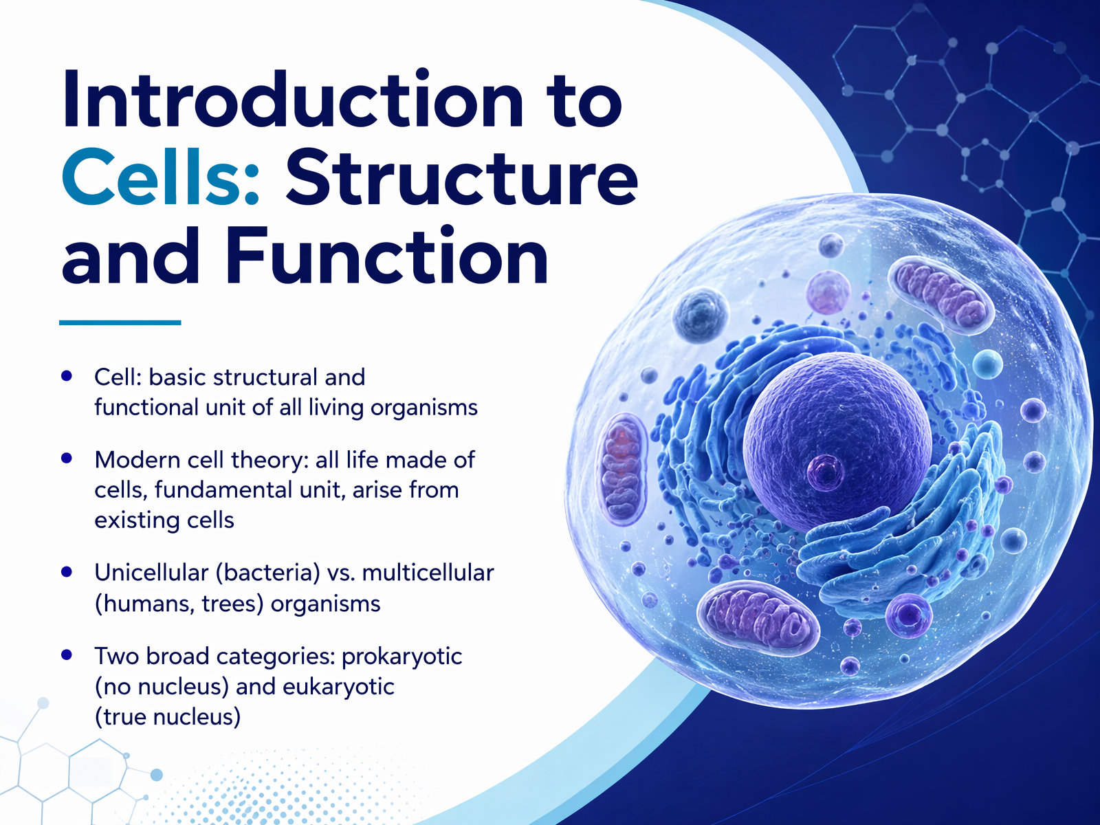
- 01Introduction to Cells: Structure and FunctionWelcome. I'm glad you're here as we begin to explore the structure and function of cells. Our goal today is to build a clear mental model of what a cell is and why it matters for how life works. Let's start with a simple definition: a cell is the basic structural and functional unit of every living organism. Think of a cell as the smallest living building block. Modern cell theory tells us three key things. First, all life is made of one or more cells. Second, the cell is the fundamental unit of life. And third, all cells arise from existing cells. Some organisms are unicellular, meaning a single cell is the whole creature, like a bacterium. Others are multicellular, like a tree or a human, where trillions of cells work together. We also group cells into two broad categories. Prokaryotic cells, like bacteria, have no true nucleus. Their DNA floats freely inside. Eukaryotic cells, like those in our own bodies, have a true nucleus that houses the DNA, like a protected control room. Now, to understand why cells are built the way they are, we need to consider a physical limit. Up next, we'll explore why size matters by looking at the surface-area-to-volume ratio.2 min
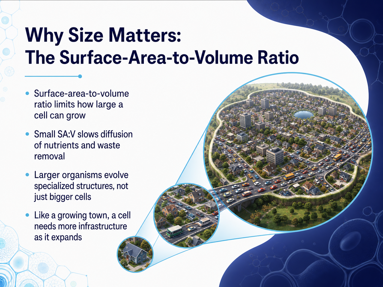
- 02Why Size Matters: The Surface-Area-to-Volume RatioBuilding on that, let's explore a major physical limit for any cell: its size. Why can't a single cell just keep growing bigger and bigger? The answer lies in something called the surface-area-to-volume ratio. Imagine a cube-shaped cell. As it grows, its volume, the space inside, increases much faster than its surface area, the outer membrane. This matters because the surface is the cell's gateway. Everything the cell needs, like nutrients and oxygen, enters through the surface. All the waste products it must get rid of exit through the surface. If the volume gets too large, the surface area can't keep up. The cell's core would be too far from the membrane, and simple diffusion becomes too slow to supply the interior or remove waste efficiently. Think of a growing town. A small village can manage with just a single main road, but a large city needs a complex infrastructure of highways, buses, and subways to move things around. Cells face the same challenge. Instead of just getting bigger, larger organisms evolve specialized transport systems, like our circulatory system, and they are built from many small cells, not one giant one. This principle of surface area to volume is a fundamental reason why life is organized the way it is. Next, let's look at the simplest living units on Earth, prokaryotic cells.
 2 min
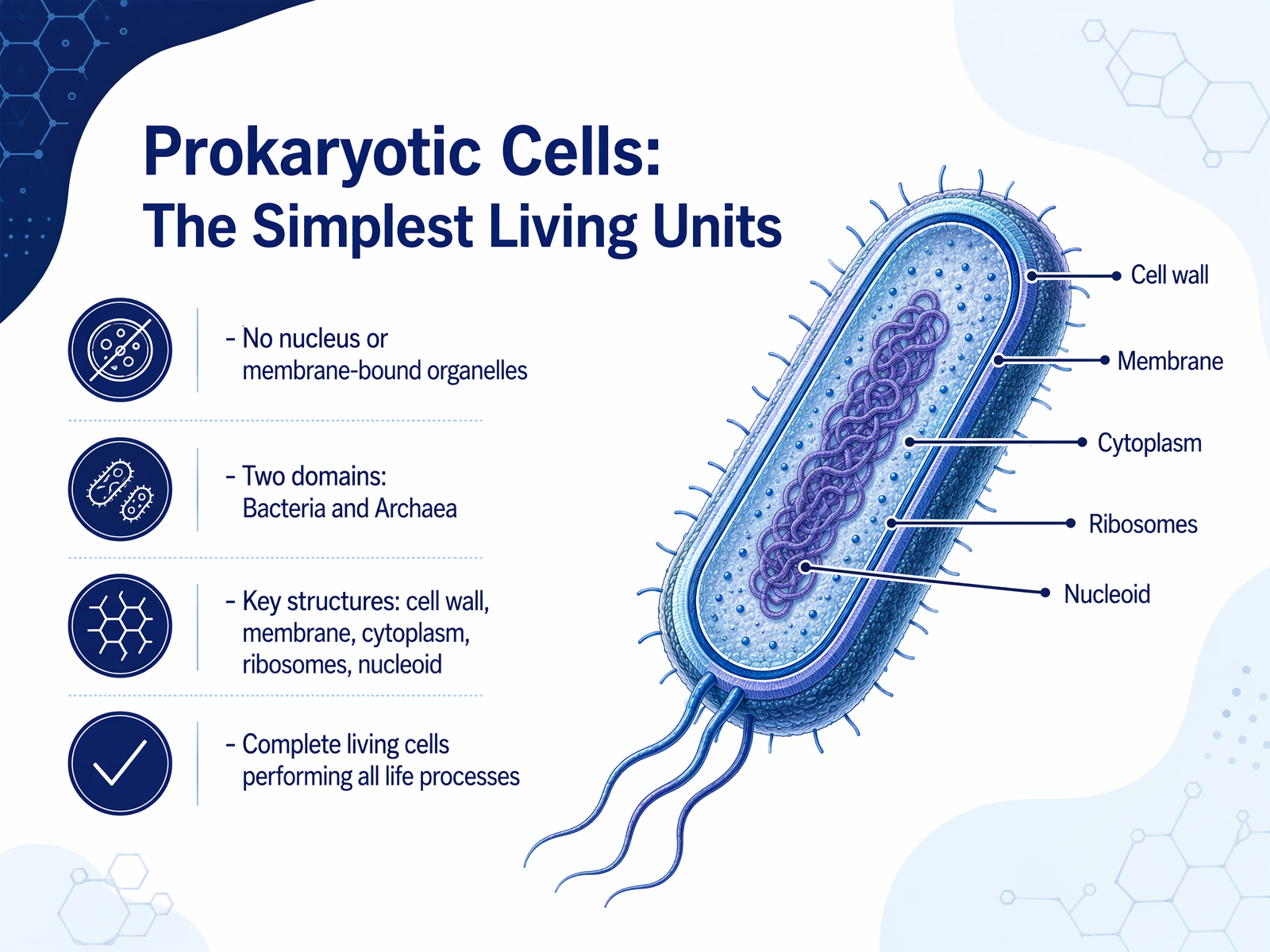
2 min - 03Prokaryotic Cells: The Simplest Living UnitsThe simplest independent living units on Earth: prokaryotic cells. The word 'prokaryotic' means 'before the nucleus,' which tells us their defining feature—they do not have a nucleus. Their DNA floats freely in a central region called the nucleoid. Prokaryotes also lack the other membrane-bound compartments you might see in more complex cells. Think of them as a compact, open-plan studio apartment, where everything happens in one main room. These cells fall into two vast domains of life: Bacteria and Archaea. While they often look similar, they are as genetically different from each other as each is from us. Despite their simplicity, a prokaryotic cell is a complete, independent living system. It has a protective cell wall, a cell membrane that controls what enters and leaves, a watery cytoplasm filling the interior, and tiny structures called ribosomes that build proteins. These cells perform all the processes of life—taking in energy, growing, and reproducing—all within a single, uncluttered package. This elegant efficiency sets the stage for the next evolutionary leap: the eukaryotic cell, where we will see the power of compartmentalization.
 2 min
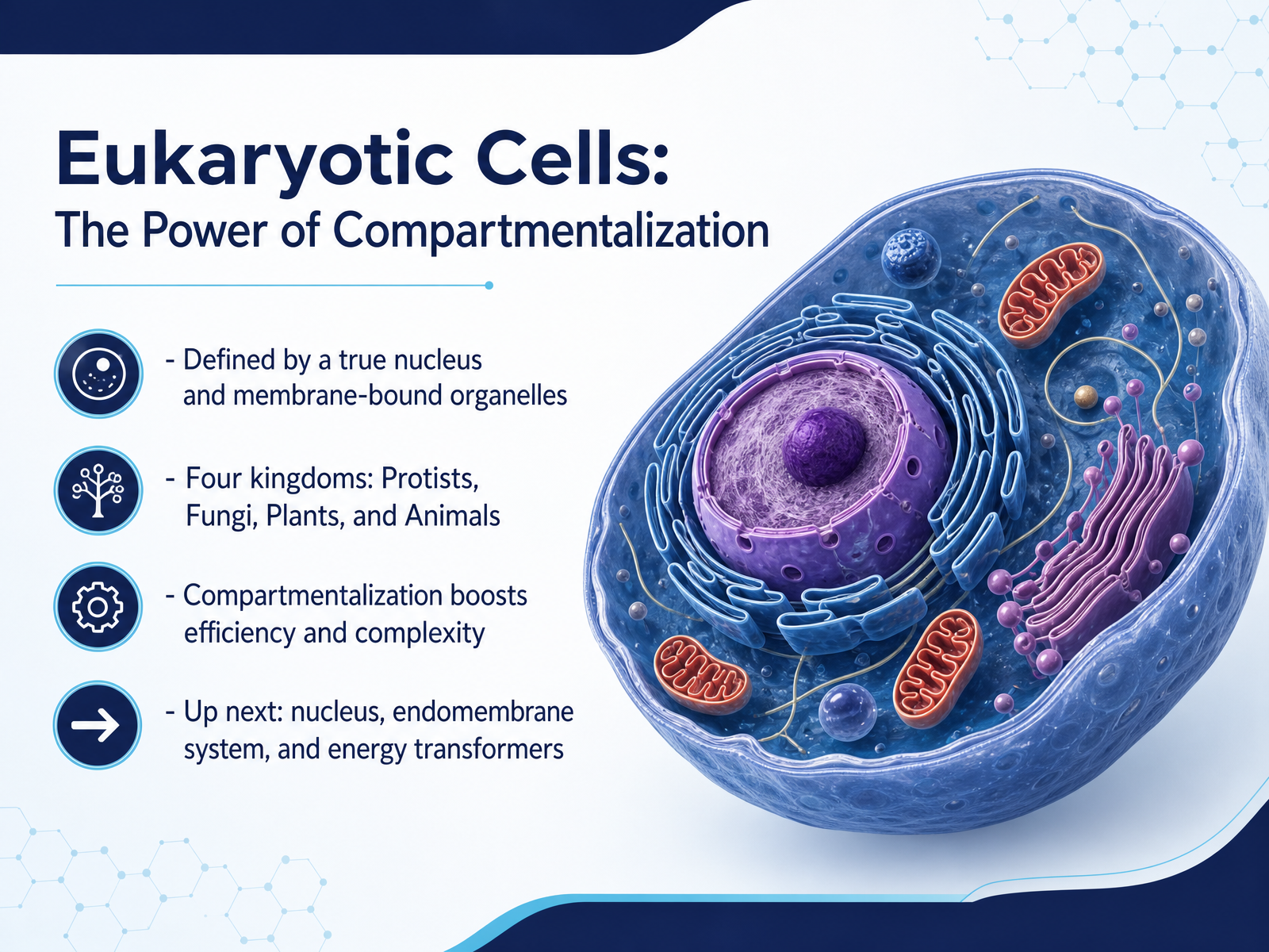
2 min - 04Eukaryotic Cells: The Power of CompartmentalizationNow let's step into a whole new level of cell design: the eukaryotic cell. The word 'eukaryote' literally means 'true kernel' or 'true nucleus,' and that's their first defining feature. Unlike bacteria, these cells keep their DNA safely tucked inside a membrane-bound control center. But the nucleus is just the beginning. Eukaryotic cells also have a variety of internal compartments called organelles, each wrapped in its own membrane. Think of the cell less like a single open room, and more like a high-tech factory with specialized rooms and workstations. This is the power of compartmentalization. It allows different jobs to happen at the same time without interfering with each other, making the cell far more efficient and complex. This design is the foundation for all the life forms we're most familiar with, grouped into four kingdoms: the Protists, a diverse collection of mostly single-celled organisms; the Fungi, like mushrooms and yeasts; the Plants; and the Animals. Each of these kingdoms is built from this same basic, compartmentalized blueprint. Over the next few minutes, we'll tour the key departments of this factory, starting with the master control center itself, the nucleus, and its protein-making partner, the ribosomes.
 2 min
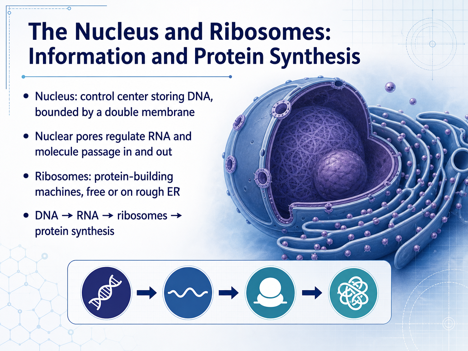
2 min - 05The Nucleus and Ribosomes: Information and Protein SynthesisNow let's zoom in on the cell's information center and its protein-building machinery. Think of the nucleus as the cell's library. It's a protected room, bounded by a double membrane, that stores the master blueprints for everything the cell does. Those blueprints are the DNA. But the DNA never leaves the library. Instead, it allows small messengers, called RNA, to carry copies of the instructions out through tiny gateways in the membrane. These gateways are the nuclear pores, and they carefully regulate what enters and exits. Once the RNA messenger exits the nucleus, it travels to a ribosome. You can picture a ribosome as a three-dimensional printer that reads the messenger's instructions and assembles a new protein, one building block at a time. Some ribosomes float freely in the cell, making proteins for internal use, while others attach to a surface called the rough ER, building proteins destined for other locations. This simple flow of information, from DNA to RNA to ribosome, is the fundamental process of protein synthesis. It's how the cell transforms stored information into the physical workers that make life happen. Next, we'll step outside the nucleus to tour the endomembrane system, a cellular factory that packages and ships these newly made proteins.
 2 min
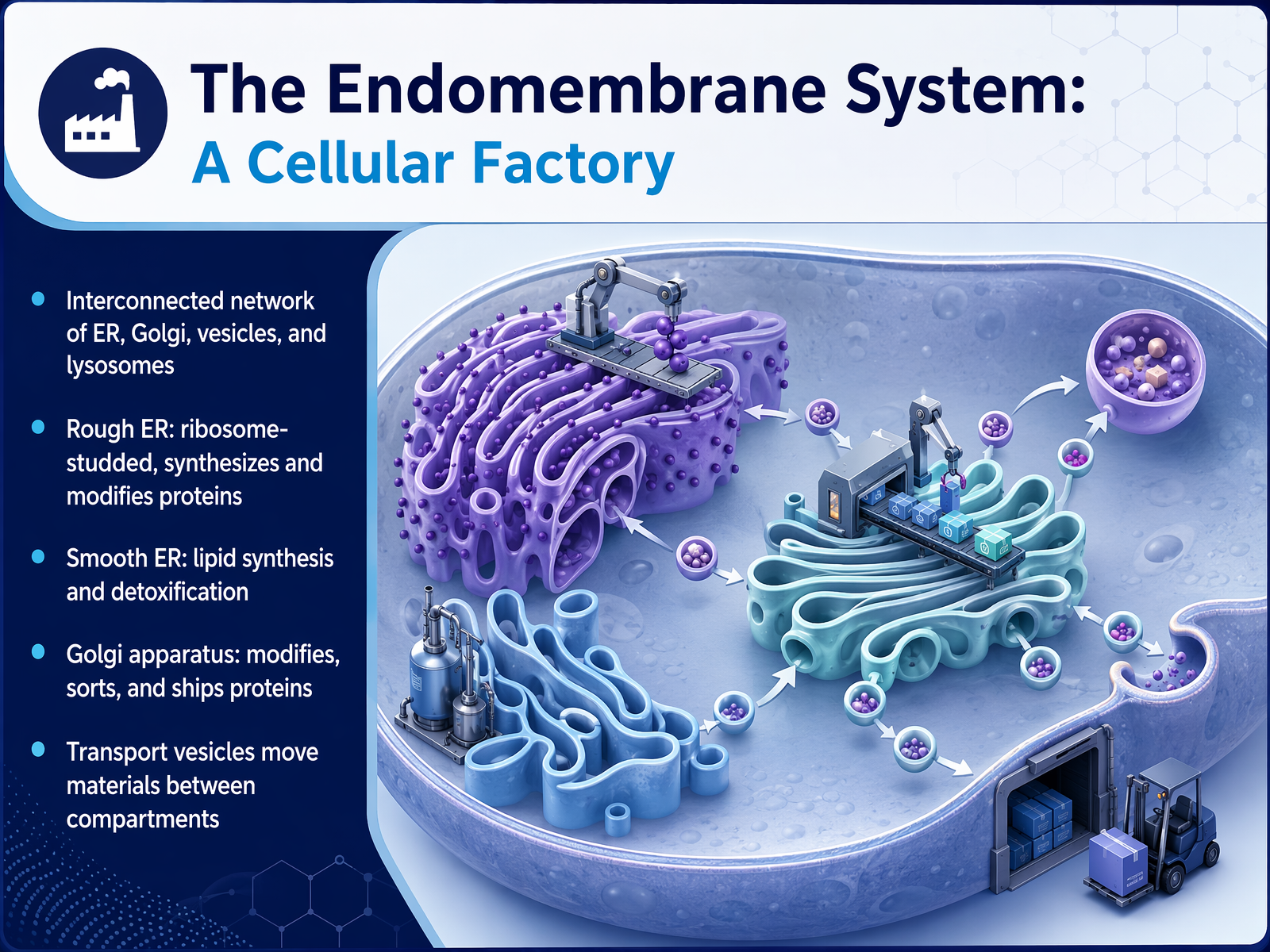
2 min - 06The Endomembrane System: A Cellular FactoryNow, let's zoom out a little and see how these parts connect into a busy cellular factory. This is called the endomembrane system. Think of it as an assembly line and shipping department inside the cell. It starts with the endoplasmic reticulum, or ER. The rough ER is dotted with ribosomes, like workers on an assembly line, where proteins are made and folded. The smooth ER is the part without ribosomes, and it handles building lipids and detoxifying harmful substances. Once a protein is ready, it gets packaged into tiny bubble-like transport vesicles. These vesicles move the cargo to the Golgi apparatus, which we can picture as the cell's sorting and shipping center. Here, proteins get final tags, telling them exactly where to go in the cell or how to be shipped out. From the Golgi, new vesicles carry the finished products to their final destinations. This entire network keeps the cell organized and efficient, making sure the right molecules get to the right place at the right time. Next, we will explore the cell's power plants, the mitochondria and chloroplasts, and see how they transform energy.
 2 min
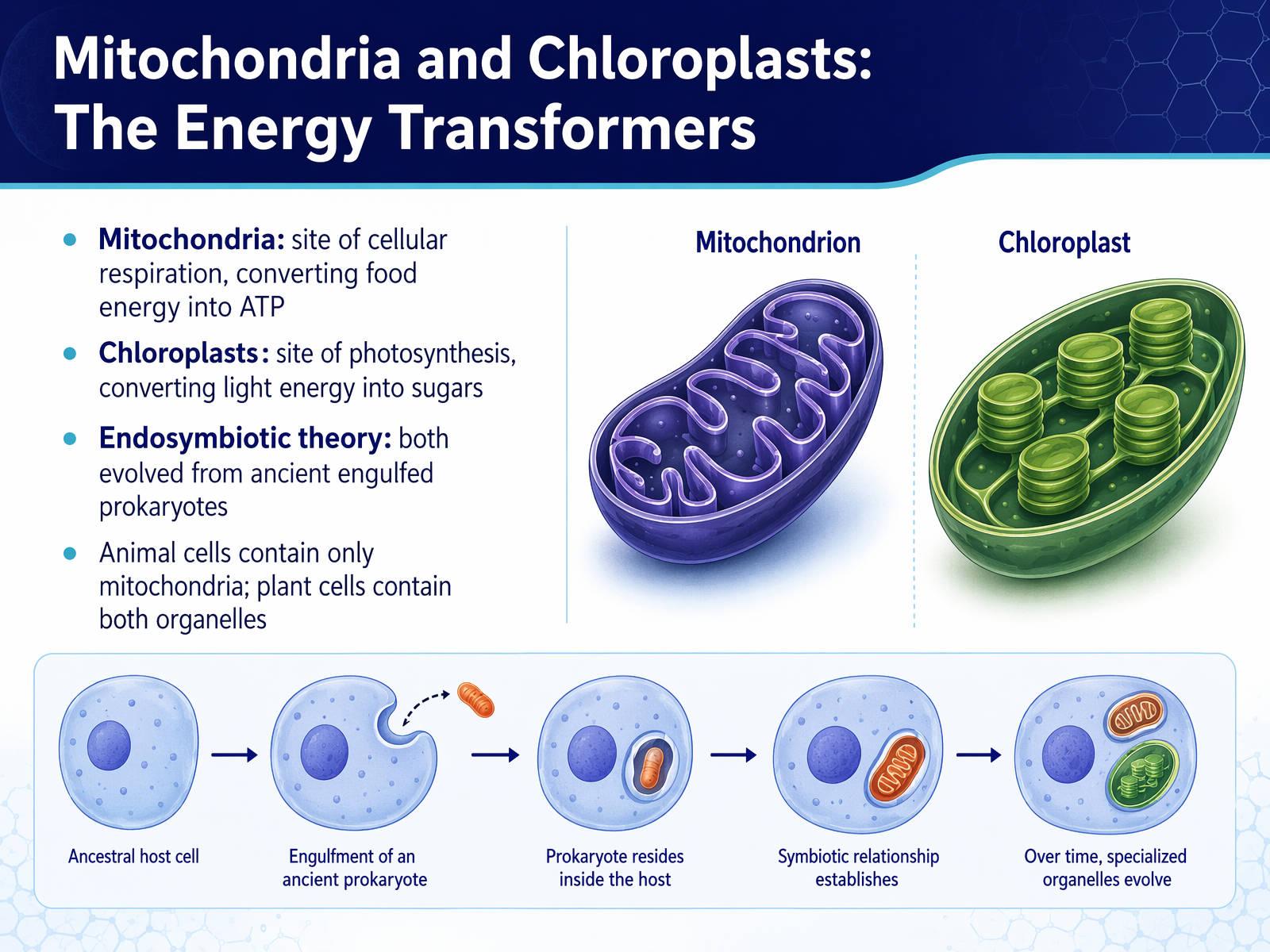
2 min - 07Mitochondria and Chloroplasts: The Energy TransformersNow, let's look at the power stations of the cell. Mitochondria and chloroplasts are specialized structures that transform energy, making it usable for life. Think of mitochondria as the cell's digestive power plants. They take the food you eat and, through a process called cellular respiration, convert that chemical energy into a molecule called ATP. ATP is the immediate energy currency that powers nearly everything your cells do. Chloroplasts, found only in plants and algae, are like tiny solar panels. They capture light energy from the sun and, through photosynthesis, lock it into the chemical bonds of sugar molecules. This sugar then feeds the plant, and eventually, feeds us. An amazing fact is that both mitochondria and chloroplasts likely evolved from ancient bacteria that were swallowed by larger cells long ago, a relationship called the endosymbiotic theory. This is why an animal cell like yours only has mitochondria, while a plant cell has both. Now, let's shift our focus from internal energy to the cell's structural framework and its boundaries.
 2 min
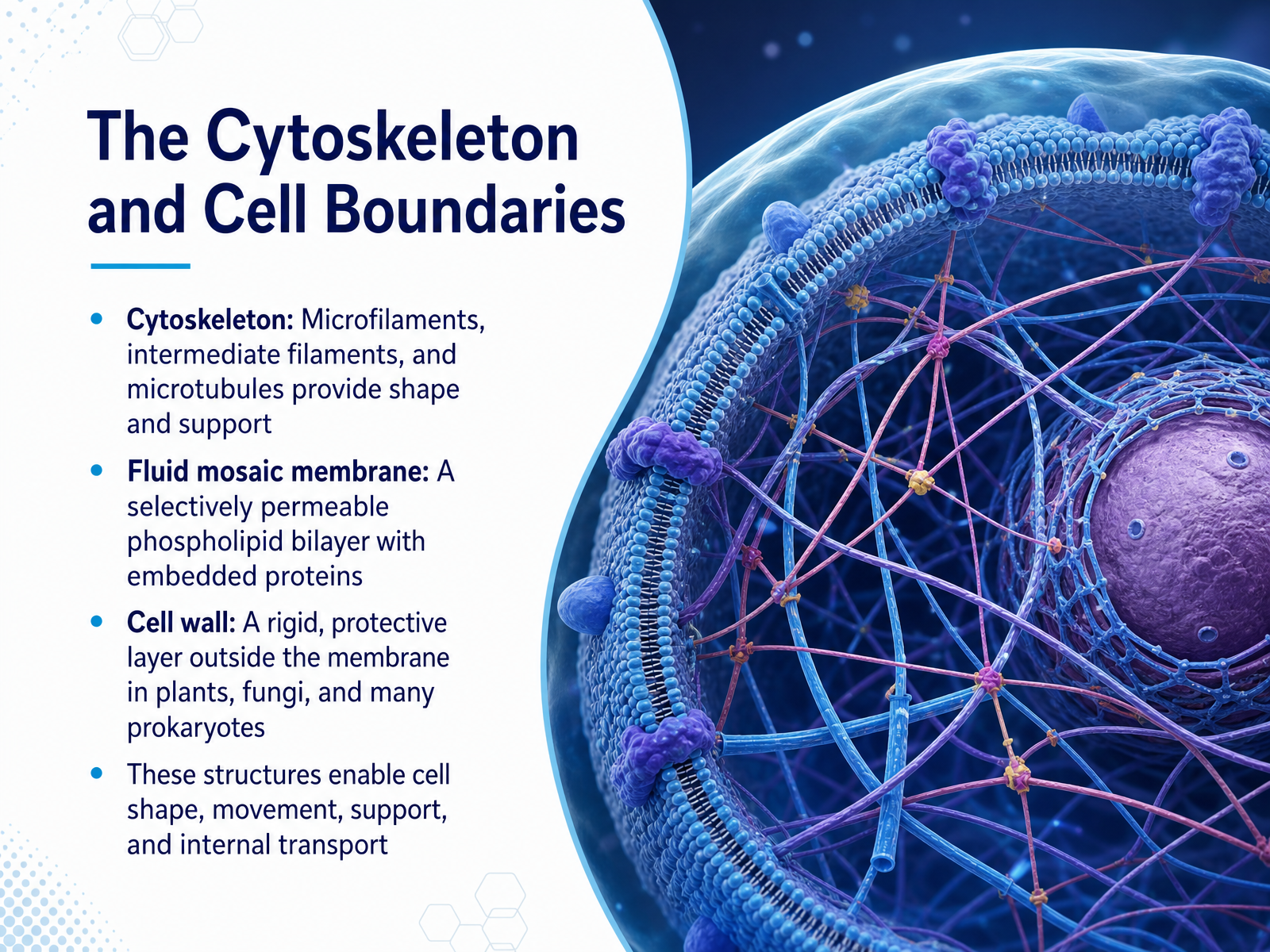
2 min - 08The Cytoskeleton and Cell BoundariesNow let's look at the cell's internal framework and its boundaries. The cytoskeleton is a network of protein fibers that gives the cell its shape, much like the poles of a tent. Microfilaments, intermediate filaments, and microtubules work together, enabling cell movement, holding organelles in place, and acting as tracks for internal transport. Surrounding the cell is the fluid mosaic membrane, a selectively permeable phospholipid bilayer with embedded proteins. Think of it as a security gate with floating guards that only lets certain things pass in and out. In plants, fungi, and many prokaryotes, a rigid cell wall sits outside this membrane, providing extra protection and support, like a strong box around a delicate package. Together, these structures maintain shape, allow movement, and control the cell's internal environment. Next, we'll compare these structures directly in plant versus animal cells.
 1 min
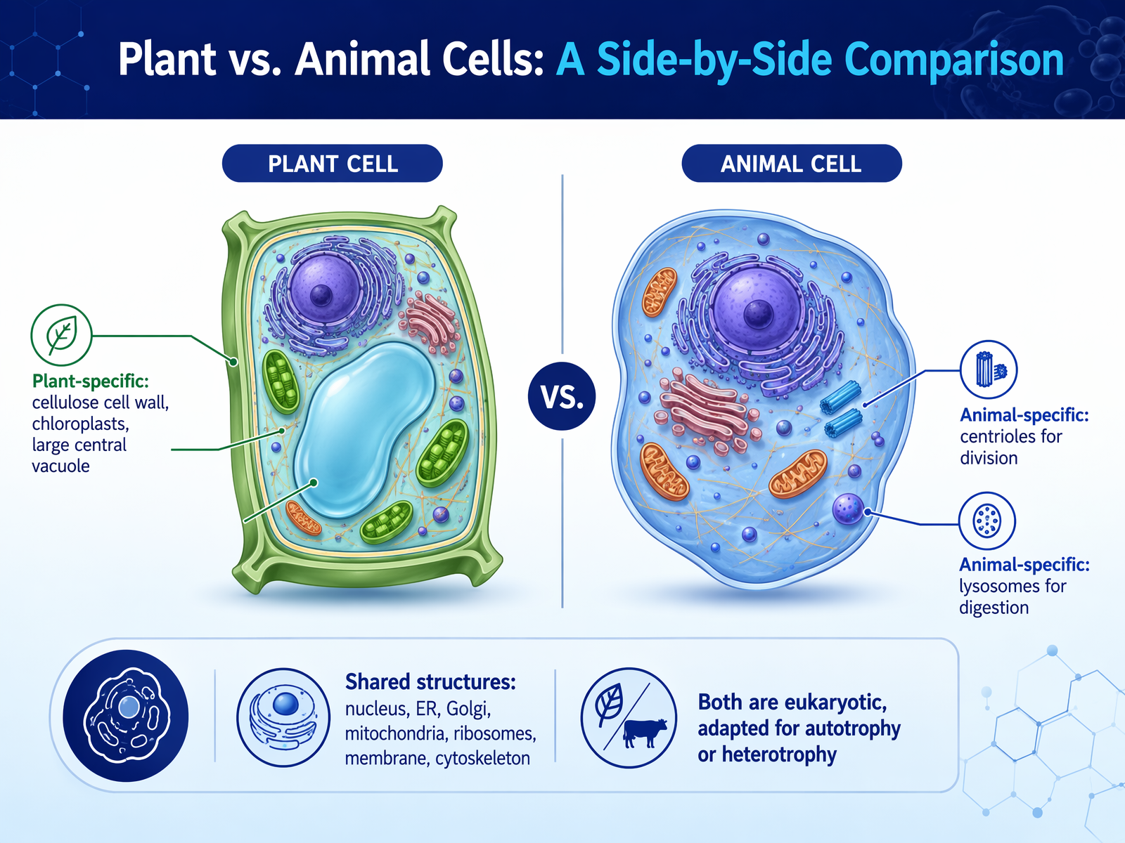
1 min - 09Plant vs. Animal Cells: A Side-by-Side ComparisonNow let's put plant and animal cells side by side. They have a lot in common, starting with the nucleus, which holds the DNA instructions. Both also build proteins with ribosomes, process them through the endoplasmic reticulum and Golgi apparatus, and power themselves with mitochondria. They share a flexible cell membrane and an internal cytoskeleton for shape and transport. The differences explain how they live. Plant cells add a rigid cellulose cell wall for structure, chloroplasts to capture light energy, and a large central vacuole for water storage and support. Animal cells skip those, but include centrioles to help organize cell division, and lysosomes—special compartments that break down waste and recycle materials. Both are eukaryotic, so their DNA is protected inside a nucleus. The key is that plants are adapted for autotrophy, making their own food, while animals are heterotrophs, taking in food from outside. Next, you'll get to see these parts yourself in a virtual microscope exploration.
 2 min
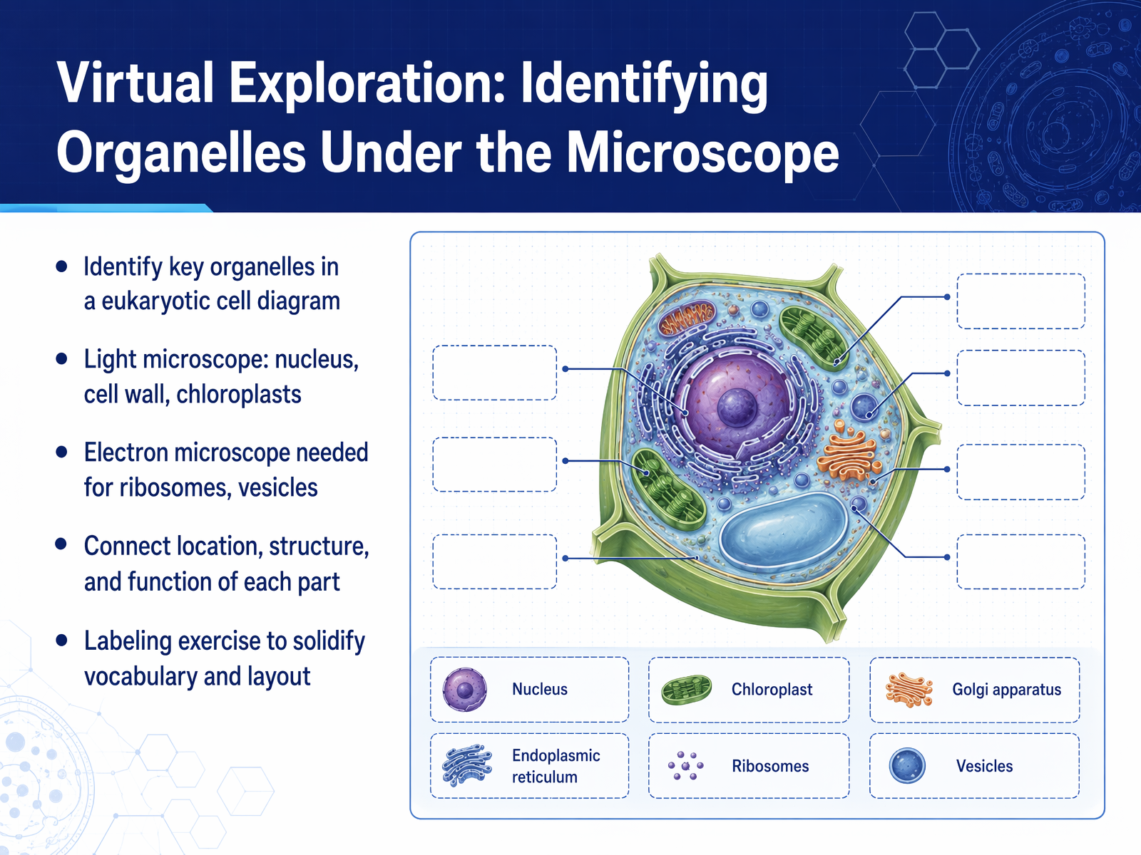
2 min - 10Virtual Exploration: Identifying Organelles Under the MicroscopeNow, let's apply what we've learned through a virtual exploration. On your screen, you'll see a diagram of a eukaryotic cell, and we're going to identify key organelles. Think of this as a tour. You'll be using a light microscope, which is like a strong magnifying glass, to spot the larger, more obvious structures. You can easily pick out the nucleus, the cell's main control center, and if it's a plant cell, the rigid cell wall and green chloroplasts will be very clear. But to see the tiny workers, like ribosomes making proteins, or small transport vesicles, you'd need a much more powerful electron microscope. Our goal is to connect what you see, its location, with what it does. Why is the nucleus in the center? Why are chloroplasts near the top of a leaf cell? As you work through the labeling exercise, don't just memorize the names. Connect each organelle's shape and position to its job. This builds your mental map of how a living cell is organized. Now that you can identify the parts, let's zoom out to see how these individual cells work together in a system.
 2 min
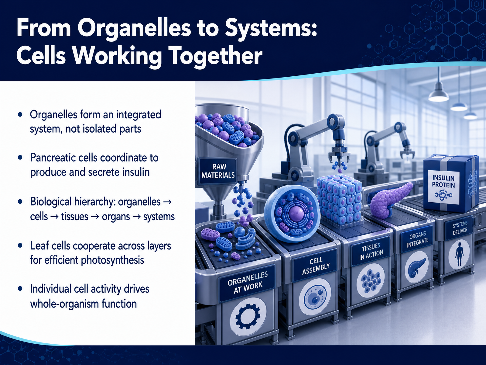
2 min - 11From Organelles to Systems: Cells Working TogetherNow let's zoom out and see how organelles work together as a system. Inside a cell, no organelle acts alone. The nucleus, ribosomes, endoplasmic reticulum, and Golgi apparatus form a connected production line, like different stations in a factory. This integrated system lets the cell respond to the body's needs. For example, in your pancreas, special cells coordinate their organelles to build and release insulin, a protein that controls blood sugar. The ribosomes assemble it, the ER folds it, the Golgi packages it, and vesicles carry it to the surface. This one cell’s activity is a tiny event, but it has a huge effect on your whole body. Now think bigger. Organelles build cells. Cells of the same type group into tissues. Tissues form organs, like a leaf or a pancreas. And organs work together in systems. In a leaf, the upper cells are packed with chloroplasts to catch light, while lower cells have air spaces to let in carbon dioxide. Each cell specializes, but they all cooperate to make sugar for the plant. This hierarchy, from a single working organelle up to a whole organism, is the foundation of life. Every breath you take and every move you make depends on countless cells, each running its own integrated system, working together. Next, we'll explore how this understanding helps us in the real world, in 'Cells in the Real World: Why This Matters.'
 2 min
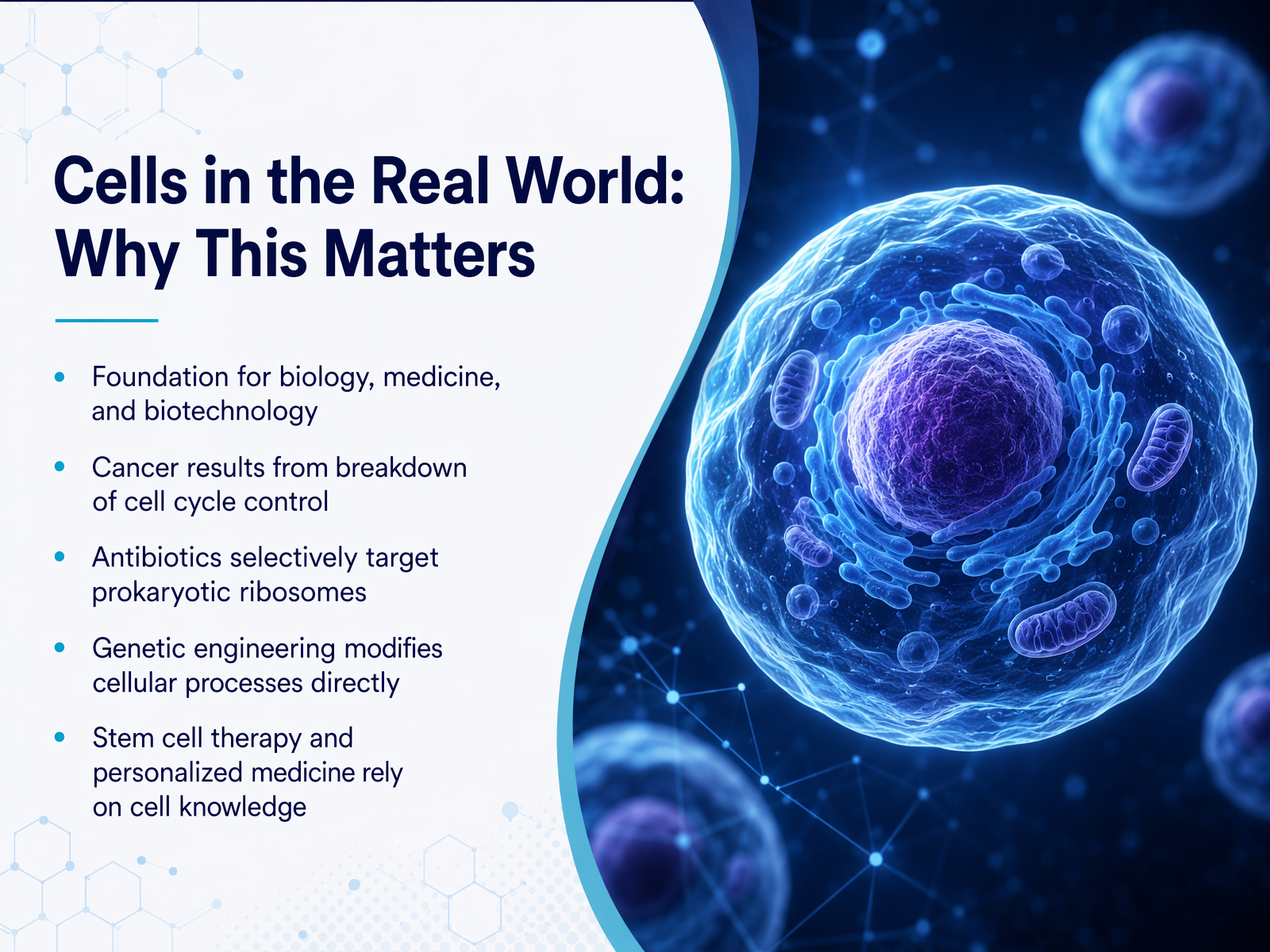
2 min - 12Cells in the Real World: Why This MattersSo why does all of this matter for how life works? Understanding cell structure and function is the foundation for nearly all of biology, medicine, and biotechnology. Every time you hear about a new medical treatment, it probably starts with our knowledge of cells. Think about cancer. Cancer is fundamentally a breakdown of the cell cycle control systems we discussed. A cell ignores the stop signals and divides uncontrollably. When we take an antibiotic for a bacterial infection, that drug works by selectively targeting prokaryotic ribosomes, which are different from our own. This is a perfect example of how a tiny structural difference can be used to save a life. Genetic engineering, from creating insulin for diabetics to developing drought-resistant crops, works by directly modifying these cellular processes we've been exploring. And the future of medicine, with stem cell therapies and personalized treatments, is completely built on our deep understanding of how cells work. You've now built the first and most important mental model for understanding the living world. Thank you for joining me, and I encourage you to keep asking questions about the amazing universe inside every cell.
 2 min
2 min
Take the deck with you
Download this course as a file — free, no sign-up needed.
- PDF handoutEvery slide page, ready to print or share.13 pages · 4.0 MBDownload
- Narrated PowerPointThe deck that presents itself — every slide carries the digital human's narration video.13 pages · 14.3 MBDownload
- PowerPoint slidesThe full deck as a .pptx — open it in PowerPoint, Keynote, or Google Slides.13 pages · 3.9 MBDownload
Free to use in your own training — please keep the PersonWise credit page at the end.
Have your own deck? Turn it into a course