Begin
14 pages · ~28 min
Diffusion and Osmosis Across Cell Membranes
This training explains diffusion and osmosis, teaching learners how substances cross cell membranes. Ideal for biology students and educators.
A digital instructor presents all 14 pages. Hold “Ask” at any point and ask out loud — the answer comes from this course. No sign-up needed.
What you’ll learn
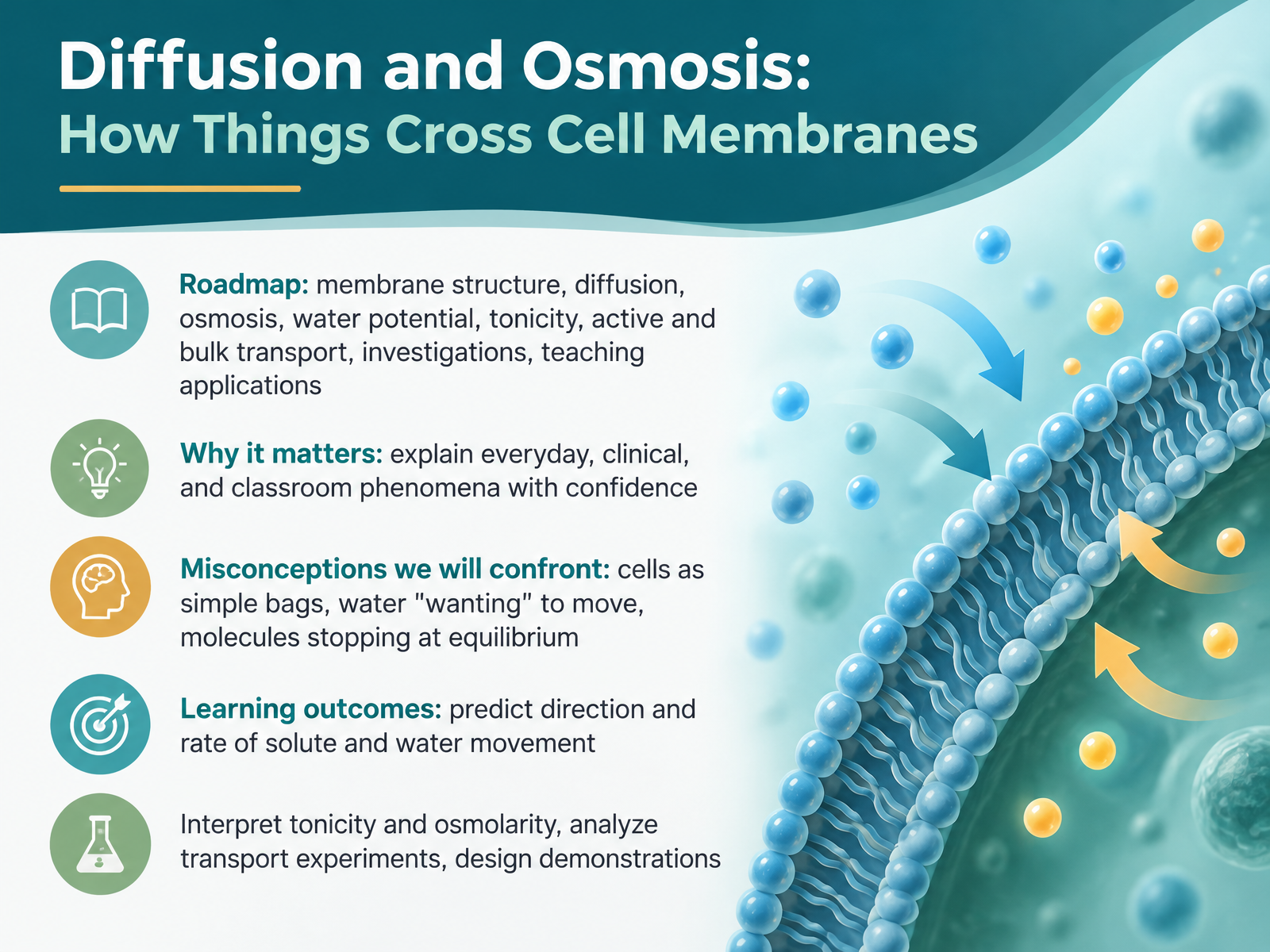
- 01Diffusion and Osmosis: How Things Cross Cell MembranesWelcome. Today we're going to explore diffusion and osmosis, two processes that explain how things cross cell membranes. Our goal is practical: by the end, you'll be able to predict the direction and rate of solute and water movement, interpret tonicity and osmolarity, and analyze transport experiments with confidence. We'll move through membrane structure, diffusion, osmosis, water potential, tonicity, active and bulk transport, then finish with investigations and teaching applications. Why does this matter? These ideas explain everyday events like a wilted salad, clinical situations like intravenous fluids, and classroom demonstrations. Along the way, we'll confront three common misconceptions: that cells are simple bags, that water somehow wants to move, and that molecules stop moving at equilibrium. Hold those in mind as we go. Let's begin with membrane structure and selective permeability.doi.orgdoi.orgtandfonline.com+22 min
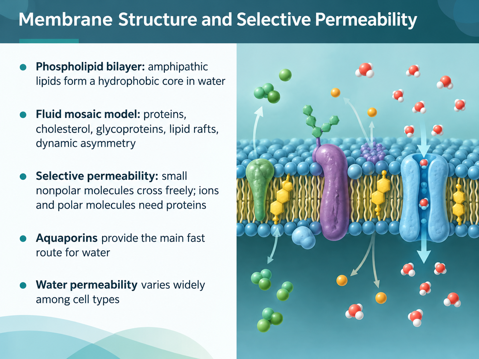
- 02Membrane Structure and Selective PermeabilityNow, before we can follow molecules across the membrane, we need to understand the barrier itself. The membrane is built from a phospholipid bilayer. Each lipid has a water-loving head and a water-fearing tail, so in water, the tails hide inward, forming a hydrophobic core. Picture a crowd turning its umbrellas toward the rain. This gives us the fluid mosaic model. Proteins, cholesterol, and glycoproteins drift within a dynamic, slightly asymmetric sea. This structure makes the membrane selectively permeable. Small nonpolar molecules, like oxygen and carbon dioxide, cross freely. Ions and most polar molecules need protein channels or carriers. Water is interesting. It crosses slowly through the lipid bilayer, but aquaporins provide the main fast route. And water permeability varies widely among cell types, depending on membrane lipids and aquaporin expression. So the membrane is not a wall; it is a controlled gateway. Next, let's look at diffusion, random motion and the power of gradients.
 pmc.ncbi.nlm.nih.govdoi.orgtandfonline.com+22 min
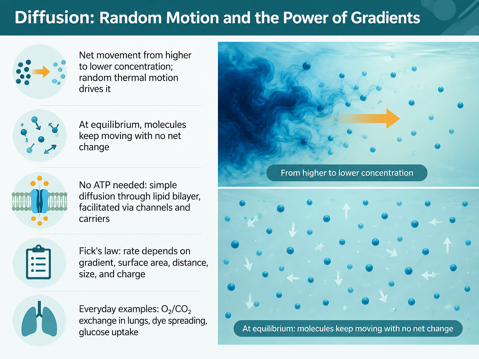
- 03Diffusion: Random Motion and the Power of GradientsNow let's look at diffusion itself. Diffusion is the net movement of particles from an area of higher concentration to an area of lower concentration, driven by random thermal motion. Picture a drop of dye in a glass of water. It spreads without any stirring until the color is even. At that point, molecules are still moving, but there is no net change. That is equilibrium. Diffusion needs no ATP. Small, nonpolar molecules like oxygen and carbon dioxide cross the lipid bilayer directly, which is simple diffusion. Ions and polar molecules need channels or carriers, which is facilitated diffusion. How fast diffusion happens is described by Fick's law. The rate depends on the concentration gradient, the surface area available, the distance across the membrane, and the size and charge of the molecule. In your lungs, oxygen and carbon dioxide exchange this way. Glucose uptake uses carrier proteins with a maximum rate. Next, we look at facilitated diffusion in more detail, including channels, carriers, and saturation.
 pmc.ncbi.nlm.nih.govbio.libretexts.orgopenstax.org+22 min
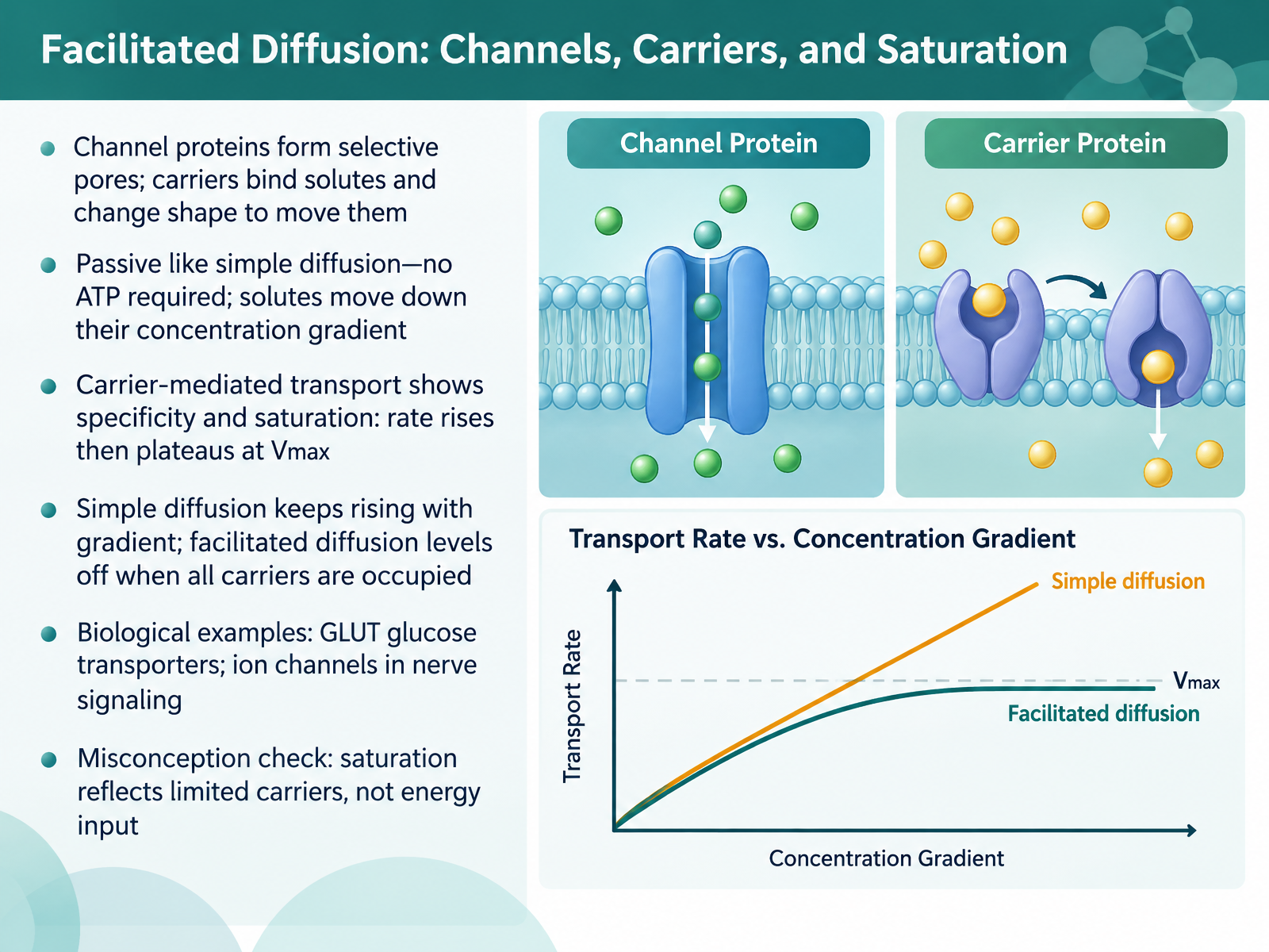
- 04Facilitated Diffusion: Channels, Carriers, and SaturationNow let's look at how larger or charged molecules get help crossing the membrane. Facilitated diffusion uses two kinds of proteins. Channel proteins form selective pores, like a dedicated doorway for one type of ion. Carrier proteins bind a solute and change shape to move it across. Both are passive, meaning no ATP is required. Solutes still move down their concentration gradient. But carriers show specificity and saturation. As the gradient steepens, the transport rate rises, then plateaus at a maximum called V max. Why? Because every carrier is occupied. Simple diffusion, by contrast, keeps rising with the gradient. Think of glucose entering cells through GLUT transporters, or ion channels firing in nerve signaling. Here's a common misconception: saturation reflects a limited number of carriers, not energy input. So the bottleneck is protein availability, not ATP. Coming up next, we turn to water movement in Osmosis and Water Potential: Beyond 'Water Wants to Move'.
 pmc.ncbi.nlm.nih.govbio.libretexts.orgopenstax.org+22 min
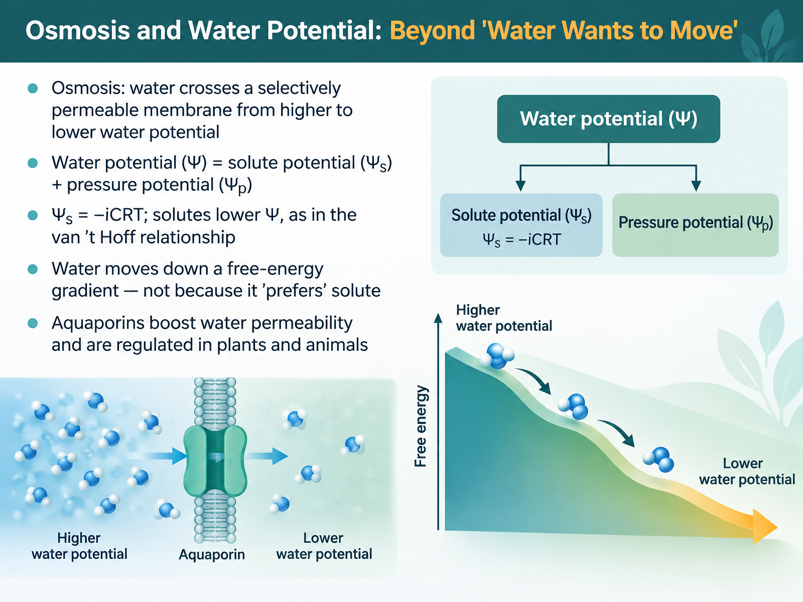
- 05Osmosis and Water Potential: Beyond 'Water Wants to Move'Let's go beyond the phrase water wants to move. Osmosis is water crossing a selectively permeable membrane from higher water potential to lower water potential. Think of water potential as the free energy of water. The formula is water potential equals solute potential plus pressure potential. Solute potential equals negative i times C times R times T, the van 't Hoff relationship. In plain terms, adding solutes lowers water potential, while positive pressure raises it. So water does not prefer solute. It simply moves down a free energy gradient. And aquaporins, the water channels, can speed that movement by orders of magnitude, and they are regulated in both plants and animals. In a classroom demo, wilted lettuce firming up in water is a clear sign of water moving into cells. So keep the mechanism in mind: energy gradients drive water, and channels control the rate. Next, we'll connect this to tonicity and osmolarity, and how they let us predict cell volume: Tonicity vs. Osmolarity: Predicting Cell Volume.
 pmc.ncbi.nlm.nih.govopenstax.orgsecure-media.collegeboard.org+22 min
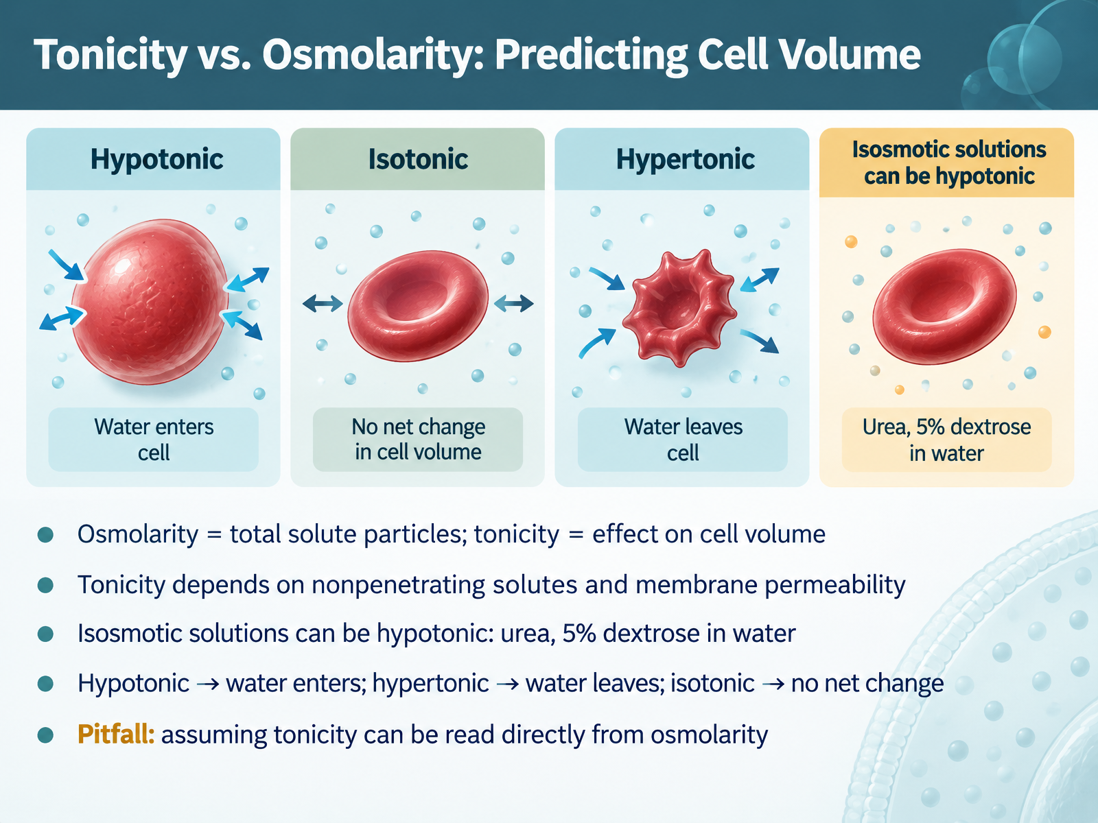
- 06Tonicity vs. Osmolarity: Predicting Cell VolumeLet's build on that and look at tonicity versus osmolarity. Osmolarity counts the total solute particles in a solution. Tonicity is different. It predicts the effect on cell volume, and it depends only on nonpenetrating solutes, meaning solutes that cannot cross the membrane. Here is the trap. A solution can be isosmotic and still be hypotonic. Think of urea, or five percent dextrose in water. Their total particle count matches the cell, but because those solutes enter the cell, water follows them in. So the rule is simple. Hypotonic means water enters. Hypertonic means water leaves. Isotonic means no net change. The pitfall is assuming you can read tonicity directly from osmolarity. You cannot. You also need to know permeability. Next, we'll see how different cells actually respond, with lysis, turgor, and plasmolysis.
 journals.physiology.orgpmc.ncbi.nlm.nih.govdoi.org+11 min
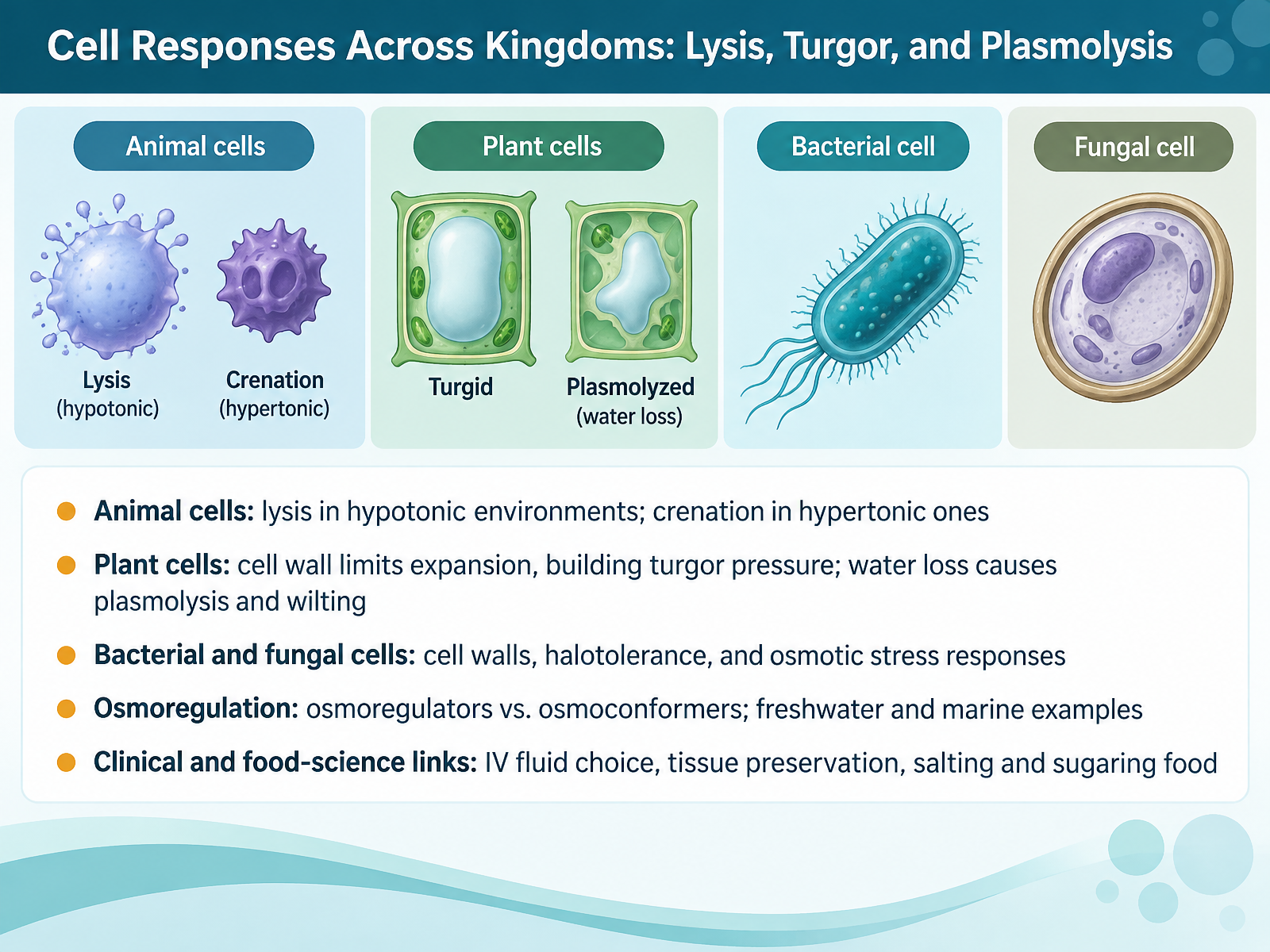
- 07Cell Responses Across Kingdoms: Lysis, Turgor, and PlasmolysisNow let's see how different cells respond to the same osmotic challenge. Animal cells have no wall, so in a hypotonic solution water rushes in and they lyse, or burst. In a hypertonic solution they lose water and crenate, shrinking and shriveling. Plant cells are protected by a rigid cell wall. Water entering builds turgor pressure, which stiffens the cell; when water leaves, the membrane pulls away from the wall in plasmolysis, and the plant wilts. Bacterial and fungal cells also have walls, and many tolerate salt through osmotic stress responses. Organisms manage this in two ways: osmoregulators actively control internal solute levels, while osmoconformers match their surroundings. You see osmosis at work clinically when IV fluids are chosen, and in food science when salt or sugar preserves tissue by drawing water out. Next, we look at active transport: pumps that work against the gradient.
 journals.physiology.orgpmc.ncbi.nlm.nih.govopenstax.org+21 min
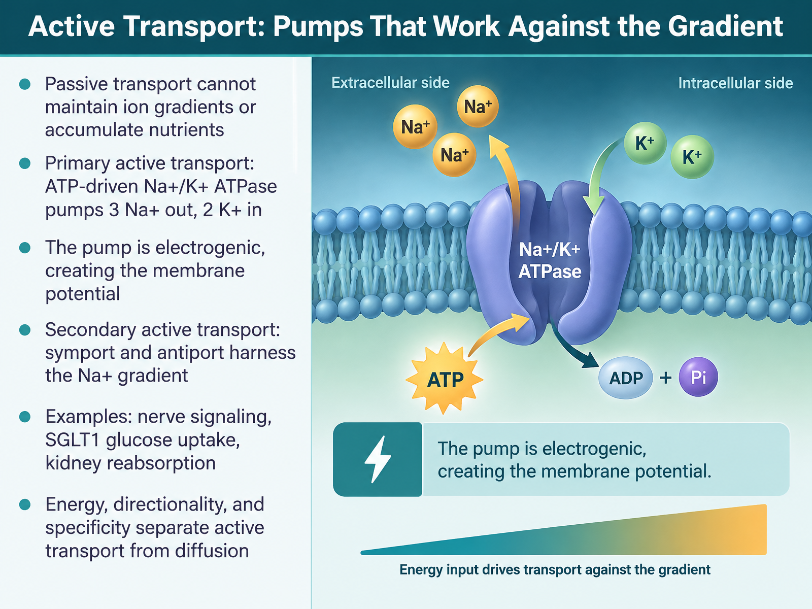
- 08Active Transport: Pumps That Work Against the GradientNow let's look at what happens when diffusion just isn't enough. Passive transport can't maintain ion gradients or stockpile nutrients. For that, cells use active transport, which spends energy to push substances against their gradient. The classic example is primary active transport: the sodium potassium pump. This pump uses ATP to move three sodium ions out of the cell and two potassium ions in. Because it moves unequal charges, it's electrogenic, meaning it helps create the membrane potential. Then there's secondary active transport, where symporters and antiporters harness that sodium gradient to move other molecules. You'll see this in nerve signaling, in SGLT1 glucose uptake, and in kidney reabsorption. So remember, energy, directionality, and specificity are what separate active transport from plain diffusion. Next, we move on to bulk transport: endocytosis and exocytosis.
 pmc.ncbi.nlm.nih.gov1 min
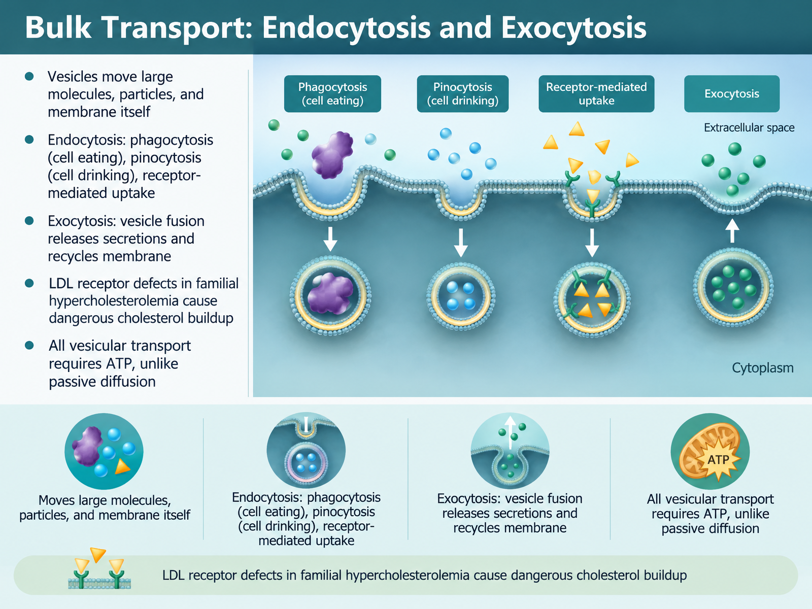
- 09Bulk Transport: Endocytosis and ExocytosisNow let's look at bulk transport. Sometimes a cell needs to move things that are simply too large for any carrier protein, so instead it uses vesicles, small membrane sacs. Moving material into the cell is endocytosis. Picture a white blood cell engulfing a bacterium, like a Pac-Man swallowing its prey. That is phagocytosis, or cell eating. Pinocytosis, cell drinking, takes in fluid with dissolved solutes. And receptor-mediated uptake is selective: receptors bind specific cargo, such as LDL cholesterol. When those LDL receptors are defective, as in familial hypercholesterolemia, cholesterol builds up in the blood to dangerous levels. Exocytosis works in reverse: vesicles fuse with the membrane and release their contents, while recycling membrane back to the surface. One key point: all vesicular transport requires energy from ATP, unlike passive diffusion. Next, we'll investigate classic experiments and data on transport.
 pmc.ncbi.nlm.nih.gov1 min
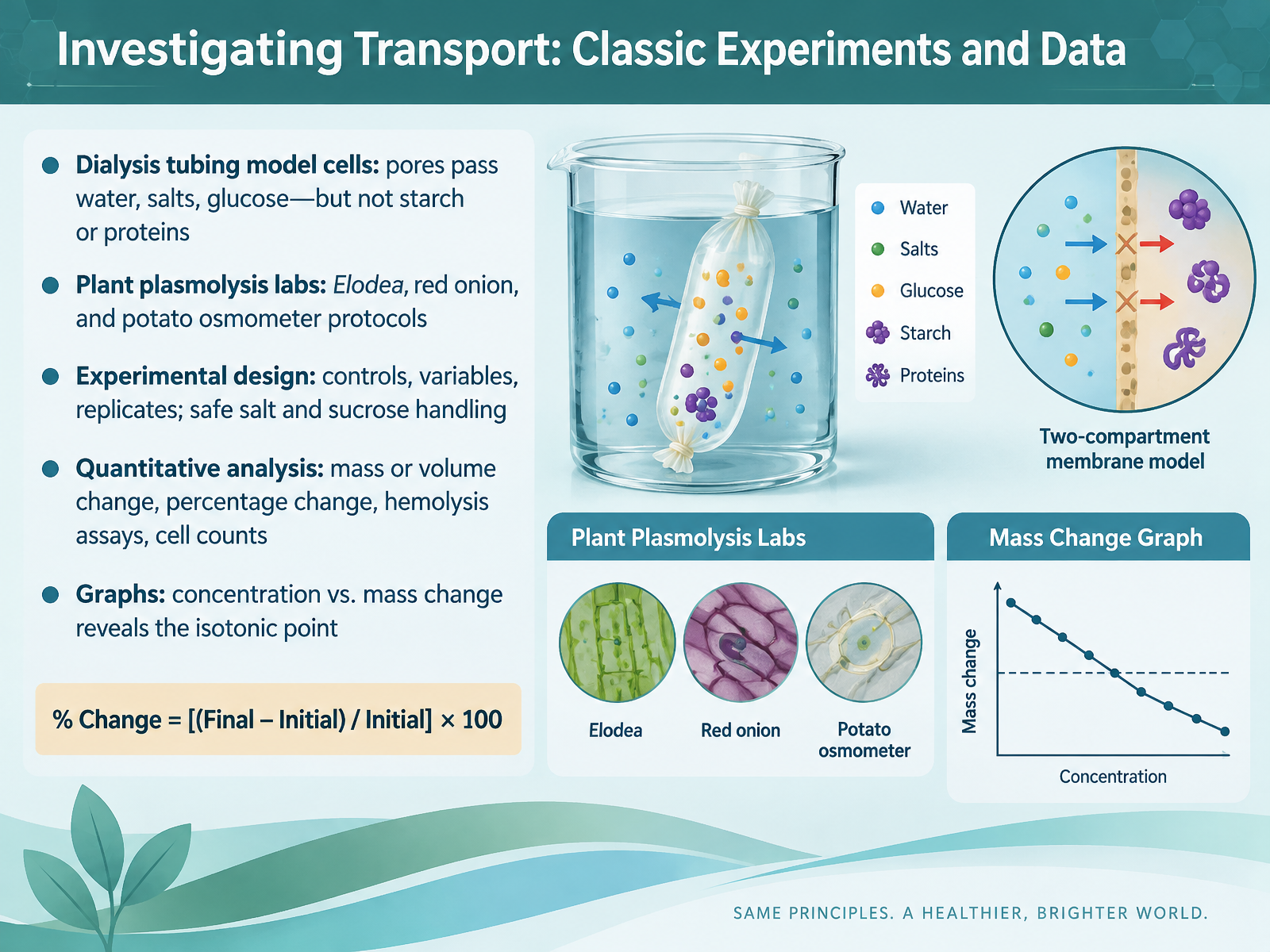
- 10Investigating Transport: Classic Experiments and DataNow let's look at how scientists investigate transport with classic experiments and real data. A common model is dialysis tubing. Its pores let water, salts, and glucose pass through, but not starch or proteins. Think of it as a sieve that only small molecules can cross. In plant plasmolysis labs, you might use Elodea, red onion, or a potato osmometer to watch water leave cells. Good experimental design matters: include controls, identify variables, and use replicates. Handle salt and sucrose safely. Then quantify results by measuring mass or volume change, percentage change, hemolysis assays, or cell counts. For example, the percentage change equals final minus initial, divided by initial, times one hundred. When you graph concentration against mass change, the point where the line crosses zero reveals the isotonic point. That is the concentration with no net water movement. Next, we'll apply these ideas to the kidney, cryopreservation, and plants.
 secure-media.collegeboard.orgjournals.physiology.orgpmc.ncbi.nlm.nih.gov+21 min
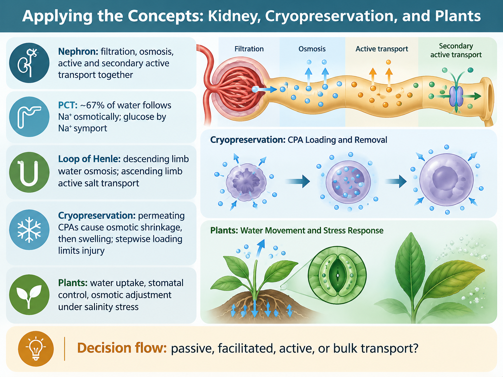
- 11Applying the Concepts: Kidney, Cryopreservation, and PlantsNow let's apply these transport mechanisms to three real systems. Start with the kidney nephron. In the proximal convoluted tubule, sodium is actively pumped out, and about sixty-seven percent of the water follows sodium by osmosis. Glucose enters with sodium through a symport protein, an example of secondary active transport. Next, the loop of Henle. In the descending limb, water leaves by osmosis. In the ascending limb, salt is moved out by active transport. Now, cryopreservation. Permeating cryoprotectants first shrink a cell osmotically, then the cell swells as the agent enters. Loading them in small steps limits injury. In plants, water uptake and stomatal control depend on osmosis, and under salinity stress, cells adjust their solute levels to keep water moving in. Here is a simple decision flow. Is the movement passive, facilitated, active, or bulk transport? Ask that question and the mechanism becomes clear. Next, we'll look at diagnosing and correcting misconceptions.
 pmc.ncbi.nlm.nih.govopenstax.orgsecure-media.collegeboard.org+22 min
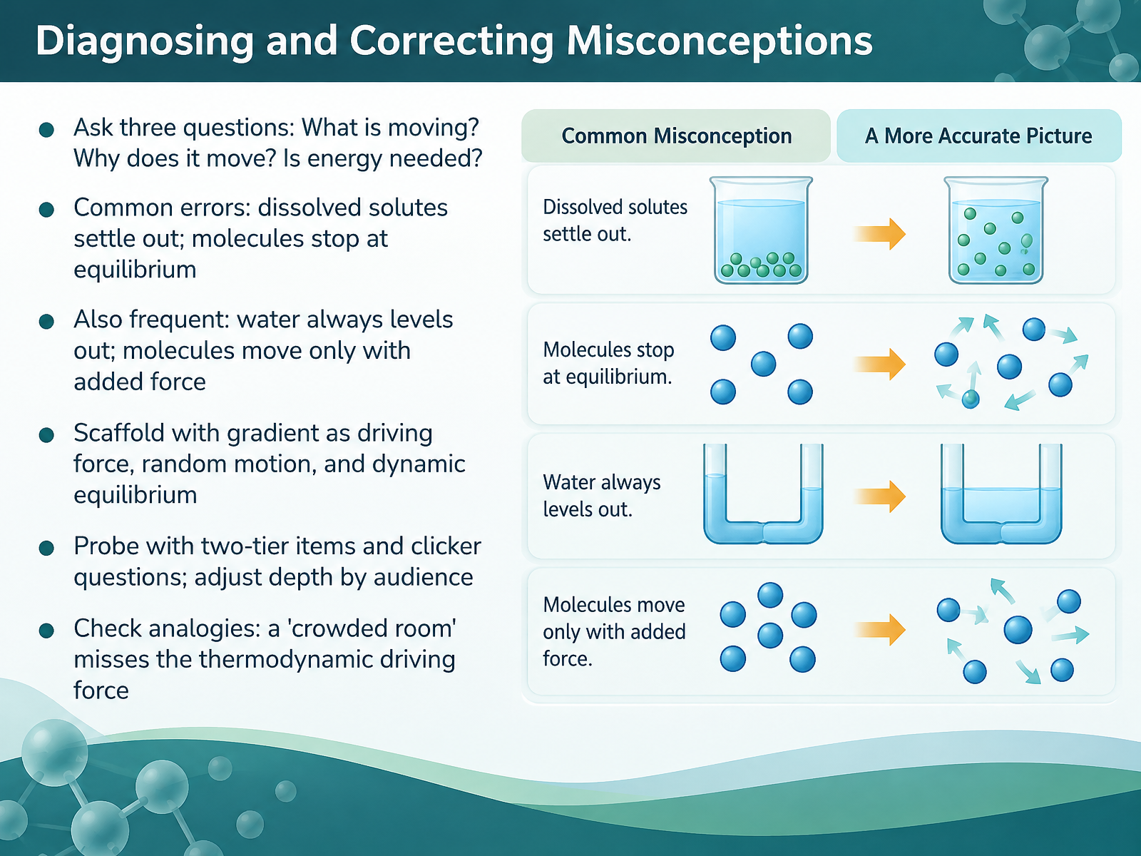
- 12Diagnosing and Correcting MisconceptionsLet's turn to diagnosing and correcting misconceptions. A simple diagnostic habit is to ask three questions about any scenario: What is moving? Why does it move? And is energy needed? Here's how errors show up in the lab, like a beaker of salt solution. Some students think dissolved solutes will eventually settle out, yet they stay randomly dispersed. Others think molecules stop at equilibrium, but equilibrium is dynamic, so molecules keep moving. Two more common errors: that water always levels out, and that molecules move only with added force. Both ignore random molecular motion. So scaffold the correct model. The concentration gradient is the driving force. Motion is random. And equilibrium is dynamic, with movement continuing in both directions. Probe with two-tier items and clicker questions, and adjust depth by audience. And check your analogies, because a crowded room picture misses the thermodynamic driving force. Next, we'll look at teaching strategies and representational sequences.
 doi.orgdoi.orgtandfonline.com+22 min
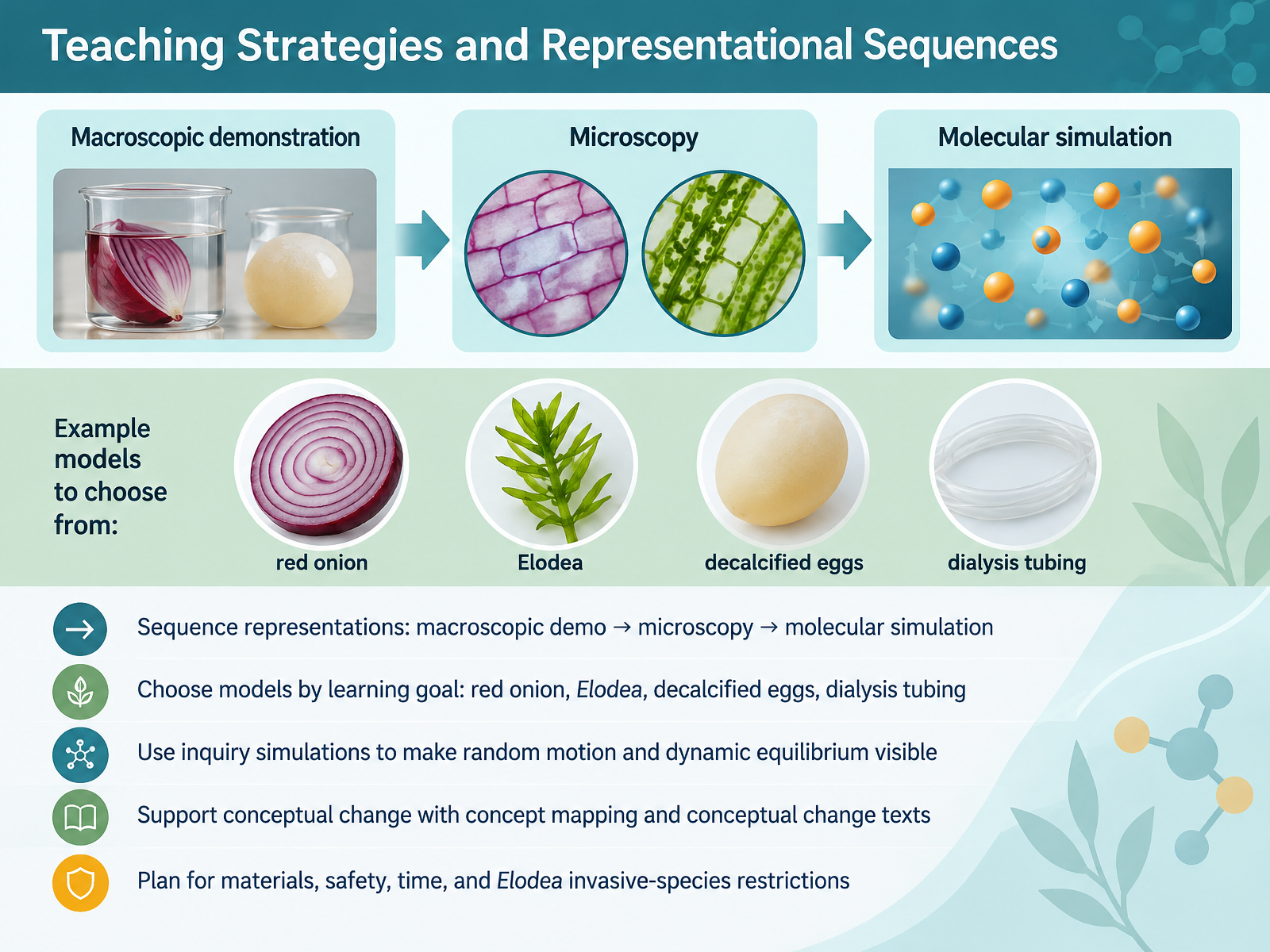
- 13Teaching Strategies and Representational SequencesLet's turn to teaching strategies and how to sequence representations. A practical order is to start with a macroscopic demonstration, move to microscopy, then finish with a molecular simulation. Choose each model by your learning goal. Red onion and Elodea show plasmolysis at the cell level, decalcified eggs and dialysis tubing model osmosis at a larger scale, and inquiry simulations make random motion and dynamic equilibrium visible. To support conceptual change, use concept mapping and conceptual change texts. Then plan the practical side: materials, safety, time, and any invasive-species restrictions on Elodea. In the next section, we'll pull this together with a summary, a self-check, and your next steps.
 pmc.ncbi.nlm.nih.govjournals.physiology.orgdoi.org+21 min
- 14Summary, Self-Check, and Next StepsLet's bring it together. We covered several key ideas: the driving forces behind diffusion and osmosis, how the membrane's selective permeability matters, water potential, and the difference between tonicity and osmolarity. Remember, tonicity is about the effect a solution has on cell volume, and it depends on nonpenetrating solutes. Now it's your turn. Try the self-check items and explain your reasoning out loud, especially on gradients, tonicity, and active transport. Then use the reflection prompts to connect these ideas to your own context. If you want to dig deeper, look at OpenStax Biology, the ODCA and DODT assessments, and research on common misconceptions. Finally, pick one action item: plan a classroom demonstration, revise an explanation you've used before, or design a two-tier question that asks students to justify their answer. Thank you for joining me. Keep asking questions and exploring how cells move substances. You've got this.
 pmc.ncbi.nlm.nih.govjournals.physiology.orgdoi.org+22 min
Take the deck with you
Download this course as a file — free, no sign-up needed.
- PDF handoutEvery slide page, ready to print or share.15 pages · 3.9 MBDownload
- Narrated PowerPointThe deck that presents itself — every slide carries the digital human's narration video.15 pages · 14.4 MBDownload
- PowerPoint slidesThe full deck as a .pptx — open it in PowerPoint, Keynote, or Google Slides.15 pages · 3.8 MBDownload
Free to use in your own training — please keep the PersonWise credit page at the end.
Have your own deck? Turn it into a course
Sources consulted
Web sources consulted while building this course.
- Development and application of a two‐tier diagnostic test measuring college biology students' understanding of diffusion and osmosis after a course of instruction — doi.org
- The Teaching and Learning of Diffusion and Osmosis: What Can We Learn from Analysis of Classroom Practices? A Case Study — doi.org
- High school students’ difficulties in understanding osmosis: International Journal of Science Education: Vol 9, No 5 — tandfonline.com
- How Effective Are Simulated Molecular-level Experiments for Teaching Diffusion and Osmosis? — pmc.ncbi.nlm.nih.gov
- High School Biology Students' Knowledge and Certainty about Diffusion and Osmosis Concepts — onlinelibrary.wiley.com
- A Brief Introduction to Some Aspects of the Fluid–Mosaic ... — pmc.ncbi.nlm.nih.gov
- Water Transport Across Cell Membranes — doi.org
- The structural basis of water permeation and proton exclusion in aquaporins (Review) — tandfonline.com
- The aquaporins - PMC — pmc.ncbi.nlm.nih.gov
- Membrane Transport — pmc.ncbi.nlm.nih.gov
- 10.2: Diffusion Across a Membrane - Passive and Facilitated Diffusion - Biology LibreTexts — bio.libretexts.org
- 5.2 Passive Transport - Biology 2e | OpenStax — openstax.org
- Simple and facilitated diffusion explained — kenhub.com
- Diffusion - Simple - Facilitated - TeachMePhysiology — teachmephysiology.com
- An update on passive transport in and out of plant cells — pmc.ncbi.nlm.nih.gov
- 30.5 Transport of Water and Solutes in Plants - Biology 2e | OpenStax — openstax.org
- BigIdea — secure-media.collegeboard.org
- Osmosis and thermodynamics explained by solute blocking — pmc.ncbi.nlm.nih.gov
- The Physical Basis of Osmosis — doi.org
- Learning (by) osmosis: an approach to teaching osmolarity and tonicity | Advances in Physiology Education | American Physiological Society — journals.physiology.org